Phase contrast is a microscopy technique that improves a contrast of unstained biological
specimens. It allows for visualizing differences in light phase caused by passing different parts of the
specimens, otherwise invisible to human's eye. This technique was developed in 1930's
by Frits Zernike,
who was awarded a Nobel Prize in 1953.
Phase contrast microscopy links:
|
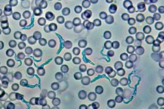 Trypanosoma gambiense, blood smear
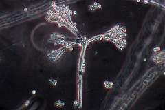 Penicillium sp.
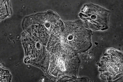 Cheek epithelium cells
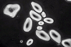 Potato starch
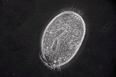 Oxytricha sp.
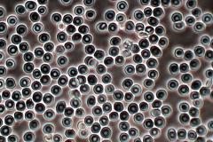 Human blood